
Page 10 / March 07
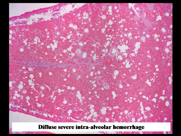
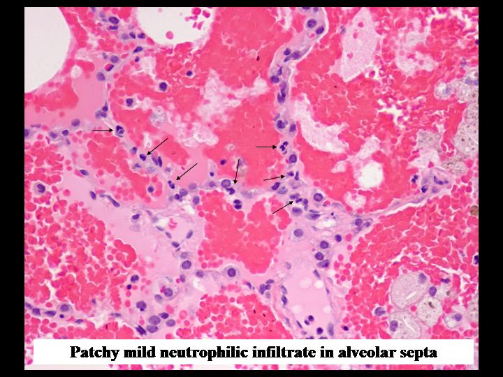
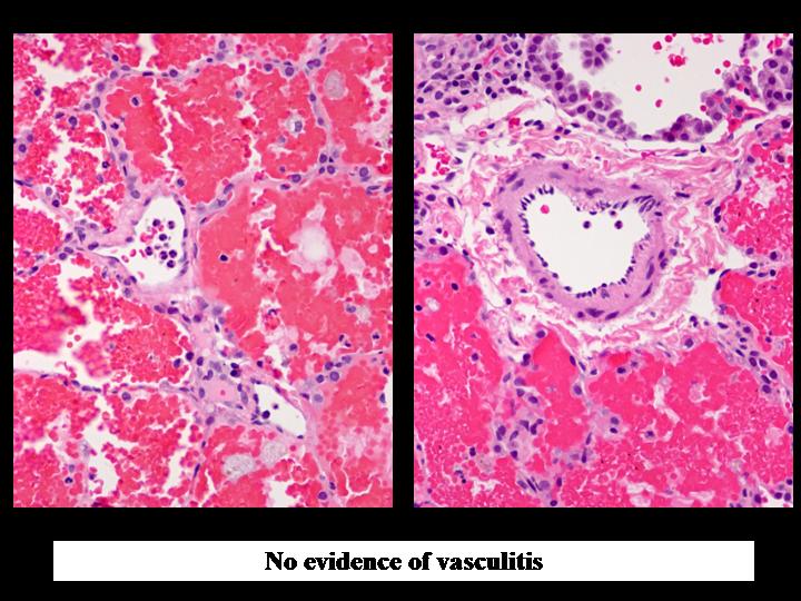
Sections show architecturally normal lung with diffuse severe intra-alveolar hemorrhage. Collections of foamy macrophages containing hemosiderin granules in their cytoplasm are present in the alveolar spaces. There are focal areas of intra-alveolar fibrin deposition, organization and fibrosis. The alveolar septae contain a mild focal infiltrate of neutrophils consistent with a mild capillaritis. Small intralobular arterioles, arteries and larger interlobular arteries show no evidence of vasculitis. The alveolar epithelial lining is intact, but shows focal type 2 pneumocyte hyperplasia. There is no evidence of hyaline membrane formation. No granulomas identified.
The rest of the pathology report was as follows:
HISTOCHEMISTRY:
Special stains for bacteria, fungi and Pneumocystis are negative.
Special stain for iron shows hemosiderin deposition in intra-alveolar foamy macrophages. Elastic trichrome stain shows preservation of the normal lung architecture, apart from the aforementioned areas of fibrosis and organization. The intrapulmonary arterioles, arteries, veins and venules are preserved and show no evidence of mural thickening, necrosis or inflammation.
IMMUNOHISTOCHEMISTRY:
Immunohistochemistry for CMV, Adenovirus, RSV, HSV-1 and -2 was negative.
IN SITU HYBRIDIZATION:
In situ hybridization for EBV is negative.
IMMUNOFLUORESCENCE:
Immunofluorescence for IgG shows discontinuous granular staining in the interstitium, most pronounced for IgG (++) but also with weak staining for IgA, IgM and fibrinogen (+) and very patchy staining for C3. The capillary basement membranes and larger vessels were negative for IgG, IgA, IgM, fibrinogen and C3.
ELECTRON MICROSCOPY:
Neutrophils are seen within capillaries and in the interstitium. Immune complexes are not identified.
 Next page /
ANSWER:
Sections show architecturally normal lung with diffuse severe intra-alveolar hemorrhage. Collections of foamy macrophages containing hemosiderin granules in their cytoplasm are present in the alveolar spaces. There are focal areas of intra-alveolar fibrin deposition, organization and fibrosis. The alveolar septae contain a mild focal infiltrate of neutrophils consistent with a mild capillaritis. Small intralobular arterioles, arteries and larger interlobular arteries show no evidence of vasculitis. The alveolar epithelial lining is intact, but shows focal type 2 pneumocyte hyperplasia. There is no evidence of hyaline membrane formation. No granulomas identified.
 |
 |
 |
| Figure 5-a | Figure 5-b | Figure 5-c |
The rest of the pathology report was as follows:
HISTOCHEMISTRY:
Special stains for bacteria, fungi and Pneumocystis are negative.
Special stain for iron shows hemosiderin deposition in intra-alveolar foamy macrophages. Elastic trichrome stain shows preservation of the normal lung architecture, apart from the aforementioned areas of fibrosis and organization. The intrapulmonary arterioles, arteries, veins and venules are preserved and show no evidence of mural thickening, necrosis or inflammation.
IMMUNOHISTOCHEMISTRY:
Immunohistochemistry for CMV, Adenovirus, RSV, HSV-1 and -2 was negative.
IN SITU HYBRIDIZATION:
In situ hybridization for EBV is negative.
IMMUNOFLUORESCENCE:
Immunofluorescence for IgG shows discontinuous granular staining in the interstitium, most pronounced for IgG (++) but also with weak staining for IgA, IgM and fibrinogen (+) and very patchy staining for C3. The capillary basement membranes and larger vessels were negative for IgG, IgA, IgM, fibrinogen and C3.
ELECTRON MICROSCOPY:
Neutrophils are seen within capillaries and in the interstitium. Immune complexes are not identified.