
Page 9 / March 07
We decided that a lung biopsy was indicated
Lung biopsy had been discussed and planned at the end of pulsing with methylprednisolone as there had been no response or at best a partial response. He was originally due for a lung biopsy on day 7 of admission but due to a combination of logistical problems in arranging PICU support and pathological laboratory examination the biopsy was done on day 10.
He had a thorascopic left lower lobe lung biopsy.
He had considerable bleeding post operatively and, though he was extubated for a few hours, he dasaturated with significant hemoptysis and had to re-intubated and ventilated. He was relatively easy to ventilate on pressure control with pressure support (PCPS) of 22/8. He was transfused.
The biopsy was an adequate specimen and the findings were reviewed jointly with the pathologists.
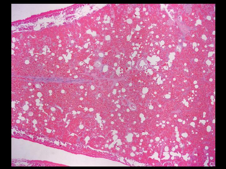
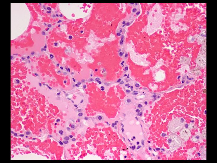
The lung biopsy was an adequate specimen comprised of a wedge of lung, 5.0 x 1.9 x 1.2 cm. The findings were jointly reviewed with the pathologists.
The next screen shows images from the lung biopsy:
 Next page /
ANSWER:
We decided that a lung biopsy was indicated
Lung biopsy had been discussed and planned at the end of pulsing with methylprednisolone as there had been no response or at best a partial response. He was originally due for a lung biopsy on day 7 of admission but due to a combination of logistical problems in arranging PICU support and pathological laboratory examination the biopsy was done on day 10.
He had a thorascopic left lower lobe lung biopsy.
He had considerable bleeding post operatively and, though he was extubated for a few hours, he dasaturated with significant hemoptysis and had to re-intubated and ventilated. He was relatively easy to ventilate on pressure control with pressure support (PCPS) of 22/8. He was transfused.
The biopsy was an adequate specimen and the findings were reviewed jointly with the pathologists.
The lung biopsy was an adequate specimen comprised of a wedge of lung, 5.0 x 1.9 x 1.2 cm. The findings were jointly reviewed with the pathologists.
The next screen shows images from the lung biopsy:
 |
 |
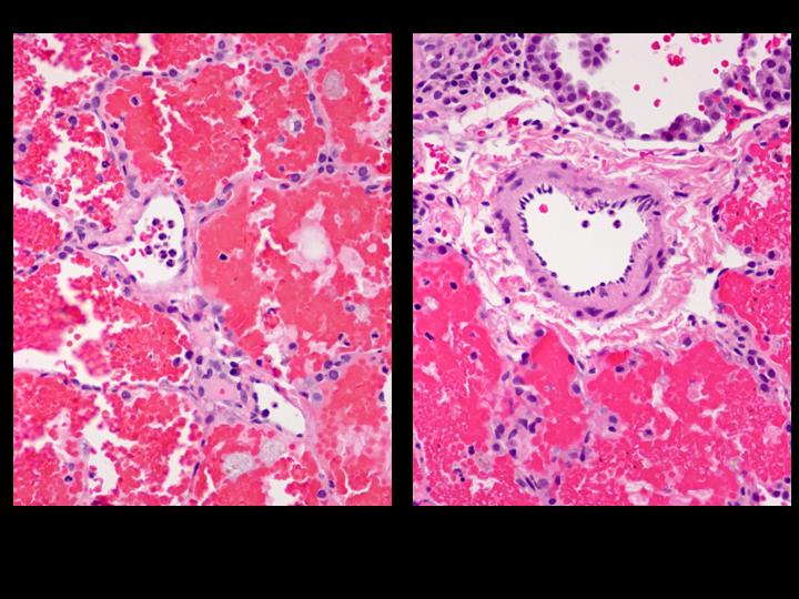 |
| Figure 4-a | Figure 4-b | Figure 4-c |