
Page 7 / May 07
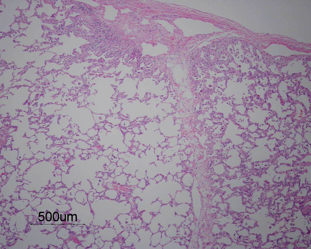
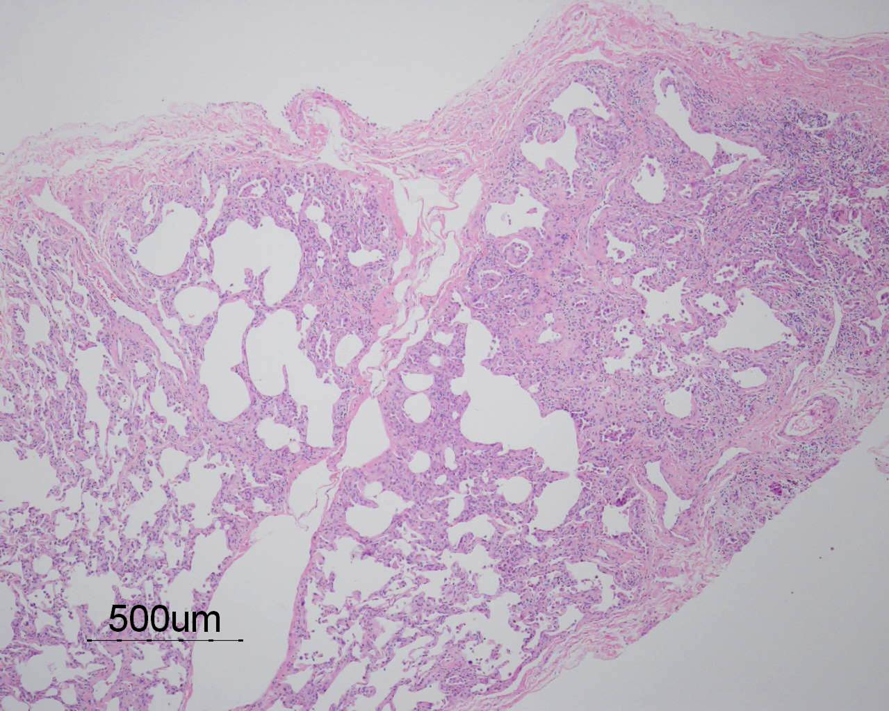
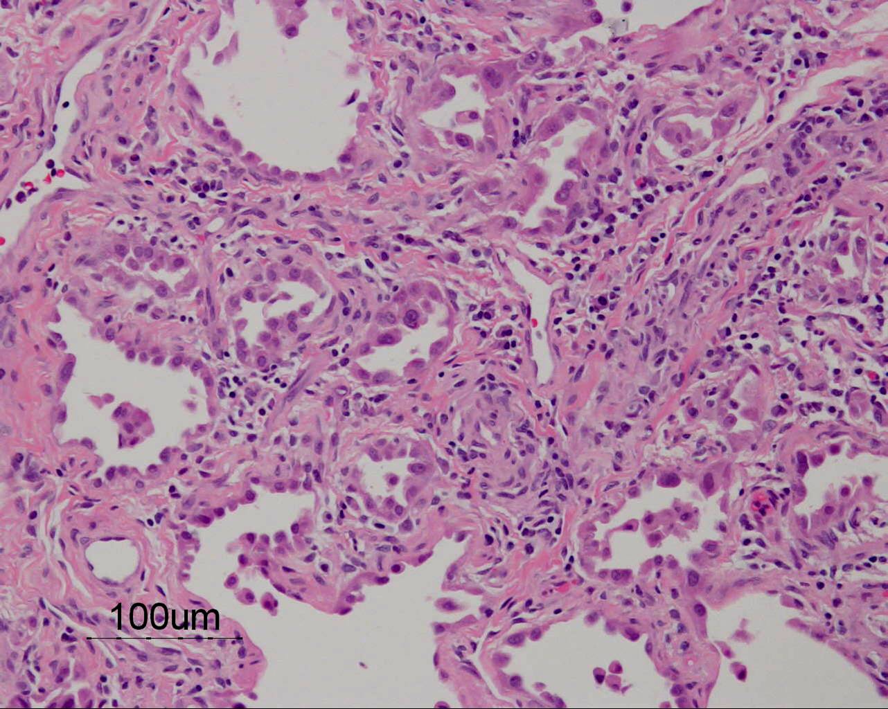
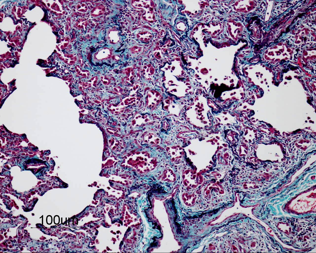
Based on the information found on the CT of the chest, LF underwent an open lung biopsy which showed usual interstitial pneumonitis (UIP) as demonstrated in the pathology below:
 Next page /
ANSWER:
- Mildly enlarged heart with prominent pulmonary vasculature.
- Air space consolidation in RLL
- Bilateral prominent reticulonodular pattern in lower zones
Based on the information found on the CT of the chest, LF underwent an open lung biopsy which showed usual interstitial pneumonitis (UIP) as demonstrated in the pathology below:
 |
 |
 |
 |