
ANSWER
At this time, our group entertained the following possibilities:1. pneumonia with pleural effusion, perhaps an empyema
2. lung abscess
3. infected peumatocele
4. diaphragmatic hernia
5. infected congenital cyst
CLINICAL PROGRESS
The girl was moderately distressed, with a respiratory rate of 60/minute, an oxygen saturation of 88%, and decreased breath sounds on left side.The
staff at the regional hospital inserted a chest tube and electively
ventilated her using low support for one night. They drained 50 ml of
pus form the cavity which was sent for Gram stain and culture. Initial
investigation included: WBC:7.3 bands: 1.3, Hb: 104 , platelets: 519,000,
and blood culture. Clindamycin and cefotaxime were initiated intravenously.
Two days later, she was afebrile, and off supplemental oxygen. The pleural
fluid culture was positive for non-typable H influenza and it was sensitive
to ceftazidime, cefuroxime, and septra. Clindamycin was discontinued
and the chest tube was removed on the 6th day. Now, after 2 weeks of
intravenous antibiotics, she was looking well and dramatically improved
clinically. Her WBC was normal and she was discharged home off antibiotics.
BELOW IS HER CHEST RADIOGRAPH ON DISCHARGE.
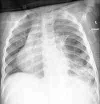
ENLARGE the chest X-ray
WHAT IS YOUR INTERPRETATION?