
ANSWER
Our Differential Diagnosis:
Diffuse Airspace Disease
–
Airspace
disease (acute)
–
Pulmonary
edema (cadiac and non-cardiac)
–
Infectious
(viral: infleunza, paqrainfluenza, RSV, CMV, adenovirus, HIV) (bacterial: mycoplasma, staph, strep, anaerobes, nocardia, MTB, NTM) (fungal: histoplasmosis and blastomycosis)
PCP
–
Neoplastic
(leukemia, lymphoma)
–
Blood
(goodpasture's, idiopathic pulmonary hemosiderosis, thrombo-embolic
disease) –
–
–
Idiopathic
(sarcoid, eosinophilic lung disease)
–
Airspace
disease (chronic)
–
Mycobacterium
tuberculosis, fungal infection, interstitial pneumonitis, lipoid pneumonia,
sarcoid, pulmonary alveolar proteinosis
DIFFUSE
INTERSTITIAL DISEASE
–
Reticulo-nodular
–
Granulomatous
- infection; mycobacterium tuberculosis versus non-tuberculous mycobacterium
–
Inhalational
exposure- organic vs inorganic
–
Idiopathic;
eosinophilic granulomatosis, sarcoid
–
Neoplastic;
leukemia
–
Hemosiderosis;
increased venous pressure
–
Repeated
hemorrhage; goodpasture's, idiopathic hemosiderosis
? Acute on chronic process (previous bronchopulmonary dysplasia)
FURTHER
HISTORY
She had a bronchoscopy and bronchoalveolar lavage in April 1998. The
lavage was cloudy, and blood-tinged in appearance. No cell count was
available. There were secretions in the RUL. Cultures were negative
for viruses, bacteria and fungal stains. Her clinical status improved
so she was discharged home on oxygen at 2 L/min with additional planned
investigations at follow-up.
At follow-up, she continued to be hypoxemic on room air. Her cardiac evaluation (echocardiogram and EKG) was normal. The interval chest xray was worse although she only required oxygen at night. Follow-up in October 1998 (6 months after presentation), she was noted to have mild clubbing on examination. Interestingly, her father was also noted to have clubbing but no respiratory symptoms.
OTHER
INVESTIGATIONS AND RESULTS
Pulmonary function tests: uninterpretable (first attempt), blood work
was normal, oximetry showed that she was hypoxemic at rest, worse with
exercise. One could argue about the need for a high resolution CT scan
to delineate the pathology further. However, it was not done at this
point.
BELOW, CHEST X-RAY from October 1998.
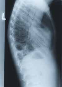
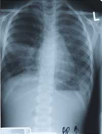
Enlarge the CHEST X-RAY LEFT, enlarge the chest x-ray RIGHT.
WHAT ARE YOUR FINDINGS. WHAT WOULD YOU DO NEXT?