
PRESENTING
FELLOW
Dr. Osama Majed
Respiratory Medicine Fellow
Albert Children's Hospital, University of Calgary
OUTLINE
–
Case
history, physical & preliminary investigations
–
Differential
Diagnosis
–
Investigations
–
Return
to the case findings & diagnosis
–
Review
of relevant background: Epidemiology, criteria for diagnosis, therapy.
CASE
OF SS
XX is a 9 month old female who was well until 3 weeks prior to admission
to the Alberta Children's Hospital (ACH). She was diagnosed to have
right upper lobe pneumonia . She was treated with oral antibiotics for
one week without improvement, and was admitted to her local hospital
and treated with intravenous cefuroxime and oral clarithromicin for
2 weeks. There was no improvement in her condition and she was transferred
to the ACH.
Review of systems revealed no chronic cough, history of diarrhea, change
in stools, nor rash. There was neither farm exposure nor sick contacts
including close contacts with anyone with a chronic cough or tuberculosis.
Past medical history showed that she was born by spontaneous vaginal
delivery. She was operated for ventricular septal defect and patent
ductus arteriosus when she was 4 months old.
Physical exam showed her to be:
–
febrile
to 39 C
–
respiratory rate of 54
–
heart rate of 165
–
blood pressure of 104/74
–
oxygen saturation of 96% on room air.
She was in moderate respiratory distress. She appeared unwell and sleepy
but awaked easily when stimulated. Positive findings on general exam
included mild sub-costal indrawing, decreased breath sounds with dullness
to percussion on the right side of the chest, and scattered crackles
diffusely with no wheeze. The liver was palpable 3 cm below the right
costal margin. First and second heart sounds were normal with an ejection
systolic murmur at left lower sternal border.
Preliminary investigations included:
–
Hb 95; MCV 71
–
Platelets 910,000
–
WBC 15.2 Neutrophils 10.5
–
Lymphocytes 2.6 Na 127
–
K4.2
–
Cl 93 ESR 47
–
PT 16
–
PTT 33.8
–
INR 1.4
The
CHEST X-RAY is seen below.
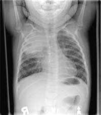
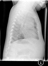
Enlarge this CHEST
X-RAY (LEFT). Enlarge this CHEST
X-RAY (RIGHT)
WHAT IS YOUR INTERPRETATION?